The cranial cruciate ligament, or CCL, in canines is analogous to anterior cruciate ligament, or ACL, in humans. The CCL is the main supporting ligament within a dog’s knee, or stifle. This ligament bears the weight of a majority of the stifle’s load, which is why instability of the CCL results in lameness, arthritis and other degenerative changes of the knee joint and surrounding tissue. The strength of this ligament will decrease as a dog ages, with larger dogs experiencing an increased rate of degeneration versus smaller breed dogs. Obese dogs are at a higher risk for CCL rupture as obesity places additional stress on the ligament. Ruptures of the cranial cruciate are the most common orthopedic injury for dogs and the leading cause for arthritic tissue in the stifle.
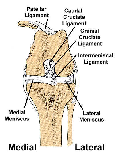
As you can see from the diagram, the cranial cruciate ligament is composed of fibers originating on the medial face of the lateral aspect of the femoral condyle, where it then inserts itself at the cranial intercondylar area of the tibia. There are two bands that make up the CCL – the craniomedial and the caudolateral. The cranial cruciate ligament receives its fresh blood supply from the synovial (joint) tissue surrounding it.
The cranial cruciate ligament’s essential function is to prevent forward movement of the tibia relative to the femur. If this forward movement is present the dog is classified as having a positive drawer test, or displaying a positive cranial drawer sign. The caudal cruciate ligament prevents backward displacement of the tibia relative to the femur, which is known as the caudal drawer. The CCL also works to limit hyperextension of the stifle and internal rotation of the tibia during flexion.
[…] Does anyone know Not sure if this shows all the ligaments. Anatomy of the Cranial Cruciate Ligament, CCL Function | Dog Knee Injury __________________ Some people are like a Slinky… not really good for anything, but you still […]
a year ago I had cruciate surgery on my left knee, 8 mo, later i had another cruciate surgery on my right knee. how common is this to have both knees have same problems, I am a 4 year old seal point registered ADBA pitt bull terrier.. someone out there please respond if we have so much in common, thank you
hi bentley,
12 weeks ago i had surgery to repair the ccl on my right leg. 4 days ago i tore the ccl on my left. at the time of the consult for the first surgery the doc recommended that both knees be done, 3 weeks apart as injury to the opposite knee is very common. my mom didn’t want to do this, but now we have to. i am a 4 year old pit bull like you.
[…] my vet, in his small consulting room, saw my dog take a few steps (leg down) and said,” Cruciate Ligament – it needs immediate surgery”. Before I had got home, he had sent an estimate of the […]